


Introduction
Materials and Methods
Sample collection and morphological observation
DNA extraction, PCR, and sequencing
Phylogenetic analysis
Results
Phylogenetic analysis
Taxonomy
Discussion
Conclusion
Introduction
Macrofungi are essential components of forest ecosystems. Of those, ectomycorrhizal (ECM) families—such as Cortinariaceae, Russulaceae, Boletaceae, and Sclerodermataceae—act as mutualistic symbionts that significantly enhance nutrient uptake and disease resistance in host trees (Smith and Read, 2008; Tedersoo et al., 2010). Although the diverse climate of South Korea supports rich fungal habitats, the national fungal inventory remains incomplete, leaving numerous taxa undocumented (Kim et al., 2023).
Identifying these fungi based solely on morphology is highly challenging due to environmental plasticity and the frequent occurrence of cryptic species complexes (Wu et al., 2014; Buyck et al., 2018). Consequently, molecular phylogenetic analysis utilizing standard DNA barcodes, such as the internal transcribed spacer (ITS) and large subunit (LSU) regions, has become indispensable for accurate taxonomic classification (Schoch et al., 2012; Vu et al., 2019). As recent molecular studies continuously uncover hidden fungal diversity across East Asia, ongoing regional surveys are crucial for building a comprehensive understanding of indigenous fungal resources (Cui et al., 2015; Park et al., 2015).
To further elucidate the macrofungal diversity of South Korea, systematic field surveys and taxonomic reviews were conducted. In this study, we report ten basidiomycetous species previously unrecorded in the country: Cortinarius acutus (Pers.) Fr., Russula burlinghamiae Singer, R. callainomarginis Jun F. Liang & J. Song, R. subatropurpurea Jing W. Li & L.H. Qiu, R. zhuzuijun Shu H. Li & X.H. Wang, Tylopilus pseudoballoui D. Chakr., K. Das & Vizzini, Pulveroboletus subrufus N.K. Zeng & Zhu L. Yang, Porphyrellus orientifumosipes Yan C. Li & Zhu L. Yang, Xerocomellus inflatus Yang Wang, B. Zhang & Yu Li, and Scleroderma venenatum Y.Zhe Zhang, C.Y. Sun & Hai J. Li. By integrating detailed morphological descriptions with phylogenetic evidence, this study updates the national inventory of indigenous biological resources and expands our understanding of their biogeographical distribution.
Materials and Methods
Sample collection and morphological observation
Fruiting bodies of the macrofungi were collected from various forest habitats in South Korea. Macroscopic characteristics, including the size, shape, color, and texture of the pileus, stipe, and hymenophore (gills or pores), were documented and photographed in the field using fresh specimens. After morphological observation, the specimens were deposited in the herbarium of the National Institute of Biological Resources (NIBR) and the Honam National Institute of Biological Resources (HNIBR). For microscopic observations, dried tissues were rehydrated and mounted in 5% potassium hydroxide (KOH) and 1% phloxine. Melzer’s reagent was used to check the amyloid or dextrinoid reactions of basidiospores. Microscopic structures such as basidia, basidiospores, and cystidia were observed and measured using an Olympus BX53 light microscope. Dimensions for basidiospores are presented as (minimum–) average (–maximum) values based on measurements of at least 30 spores per specimen.
DNA extraction, PCR, and sequencing
Total genomic DNA was extracted from the dried fruiting bodies using the DNeasy Plant Mini Kit (Qiagen, Germany), following the manufacturer’s instructions. The ITS region and the LSU of the ribosomal RNA gene were amplified using the primer pairs ITS5/ITS4 (White et al., 1990) and LR0R/LR5 (Vilgalys and Hester, 1990), respectively. Polymerase chain reaction (PCR) amplifications were performed in a C1000 Thermal Cycler (Bio-Rad, USA) under standard thermal cycling conditions. The PCR cycling conditions consisted of an initial denaturation at 95°C for 5 min, followed by 35 cycles of denaturation at 95°C for 30 s, annealing at 55°C for 30 s, and extension at 72°C for 1 min, with a final extension at 72°C for 10 min. Successful PCR amplification was confirmed by 1.5% agarose gel electrophoresis prior to sequencing. The successful PCR products were purified and sequenced by Macrogen Inc. (Seoul, South Korea).
Phylogenetic analysis
The newly generated ITS and LSU sequences were assembled and edited using SeqMan Pro (DNASTAR, USA). To determine the preliminary taxonomic placement of the specimens, the sequences were subjected to BLASTn searches (Altschul et al., 1990) against the NCBI GenBank database (NCBI, 1988). For phylogenetic tree construction, reference sequences of closely related taxa and outgroups were retrieved from GenBank. The ITS and LSU datasets were aligned individually using MAFFT v7.0 (Katoh and Standley, 2013) and manually trimmed to remove ambiguous regions. Although recent formal descriptions of complex taxa within genera such as Russula, Tylopilus, and Xerocomellus frequently employ multi-locus phylogenetic analyses (e.g., ITS, LSU, tef1, rpb2), single-locus barcodes were utilized in the present study. The ITS region is universally recognized as the primary DNA barcode for fungi and provides sufficient species-level resolution for identifying established Russula species (Schoch et al., 2012). Similarly, the LSU region is highly informative and widely accepted as a robust marker for distinguishing species within the Boletaceae, including Tylopilus and Xerocomellus (Wu et al., 2014; Vu et al., 2019). Because the objective of this study is to report previously unrecorded species in South Korea rather than to delineate novel taxa, we concluded that single-locus phylogenies—when rigorously cross-validated with macro- and microscopic morphological congruence—provide adequate and reliable resolution for accurate species identification. Phylogenetic analyses were conducted separately for each locus using the maximum likelihood (ML) method implemented in MEGA 11 (Tamura et al., 2021). The best-fit nucleotide substitution model was estimated prior to the analysis, and branch support was evaluated using 1,000 bootstrap replicates, with bootstrap values ≥ 70% displayed on the nodes of the phylogenetic trees. The generated sequences from this study were deposited in the NCBI GenBank database under the accession numbers [PZ676719 - PZ676724 for ITS; PZ677176 - PZ677185 for LSU].
Results
Phylogenetic analysis
To confirm the taxonomic placement of the ten newly recorded species, phylogenetic analyses were conducted using either ITS or LSU sequences, depending on the target taxonomic group (Figs. 1, 2, 3, 4, 5, 6, 7, 8, 9, 10).
Phylogenetic analysis based on the ITS region. The ITS region was utilized for the identification of Cortinarius acutus, the four Russula species (R. burlinghamiae, R. callainomarginis, R. subatropurpurea, and R. zhuzuijun), and Scleroderma venenatum. The ML trees generated from the ITS datasets confirmed that the Korean specimens of these taxa clustered tightly with their respective type or representative sequences. Each species formed a well-supported monophyletic group within the Cortinariaceae, Russulaceae, and Sclerodermataceae clades, respectively.
Phylogenetic analysis based on the LSU region. For the species belonging to Boletaceae—Porphyrellus orientifumosipes, Pulveroboletus subrufus, Tylopilus pseudoballoui, and Xerocomellus inflatus—phylogenetic placement was confirmed based on LSU sequences. The LSU-based ML analysis demonstrated that the Korean isolates nested securely within their respective generic clades. The specimens clustered accurately with the reference taxa for each species with strong statistical support.
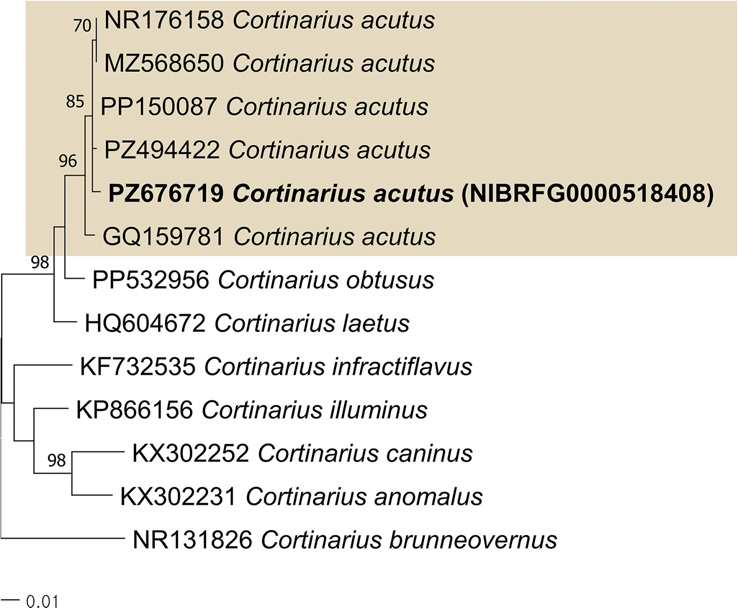
Fig. 1.
Maximum likelihood phylogenetic tree showing the phylogenetic position of Cortinarius acutus based on the internal transcribed spacer (ITS) region sequences. The species described here is highlighted with coloured blocks and bold font. The scale bar shows the expected number of nucleotide substitutions per site.

Fig. 2.
Maximum likelihood phylogenetic tree showing the phylogenetic position of Porphyrellus orientifumosipes based on the large subunit (LSU) region sequences. The species described here is highlighted with coloured blocks and bold font. The scale bar shows the expected number of nucleotide substitutions per site.

Fig. 3.
Maximum likelihood phylogenetic tree showing the phylogenetic position of Pulveroboletus subrufus based on the large subunit (LSU) region sequences. The species described here is highlighted with coloured blocks and bold font. The scale bar shows the expected number of nucleotide substitutions per site.

Fig. 4.
Maximum likelihood phylogenetic tree showing the phylogenetic position of Russula burlinghamiae based on the internal transcribed spacer (ITS) region sequences. The species described here is highlighted with coloured blocks and bold font. The scale bar shows the expected number of nucleotide substitutions per site.

Fig. 5.
Maximum likelihood phylogenetic tree showing the phylogenetic position of Russula callainomarginis based on the internal transcribed spacer (ITS) region sequences. The species described here is highlighted with coloured blocks and bold font. The scale bar shows the expected number of nucleotide substitutions per site.

Fig. 6.
Maximum likelihood phylogenetic tree showing the phylogenetic position of Russula subatropurpurea based on the internal transcribed spacer (ITS) region sequences. The species described here is highlighted with coloured blocks and bold font. The scale bar shows the expected number of nucleotide substitutions per site.

Fig. 7.
Maximum likelihood phylogenetic tree showing the phylogenetic position of Russula zhuzuijun based on the internal transcribed spacer (ITS) region sequences. The species described here is highlighted with coloured blocks and bold font. The scale bar shows the expected number of nucleotide substitutions per site.

Fig. 8.
Maximum likelihood phylogenetic tree showing the phylogenetic position of Scleroderma venenatum based on the internal transcribed spacer (ITS) region sequences. The species described here is highlighted with coloured blocks and bold font. The scale bar shows the expected number of nucleotide substitutions per site.

Fig. 9.
Maximum likelihood phylogenetic tree showing the phylogenetic position of Tylopilus pseudoballoui based on the large subunit (LSU) region sequences. The species described here is highlighted with coloured blocks and bold font. The scale bar shows the expected number of nucleotide substitutions per site.

Fig. 10.
Maximum likelihood phylogenetic tree showing the phylogenetic position of Xerocomellus inflatus based on the large subunit (LSU) region sequences. The species described here is highlighted with coloured blocks and bold font. The scale bar shows the expected number of nucleotide substitutions per site.
Taxonomy
Cortinarius acutus (Pers.) Fr., Epicrisis Systematis Mycologici: 314 (1838)
Basidiomata small to medium-sized. Pileus 10 - 20 mm in diam., initially conical to sharply conical when young, becoming campanulate and eventually nearly plane when old, consistently bearing a prominent acute umbo; upper-surface smooth, covered with whitish fibrillose veil remnants when young, becoming glabrescent when old; light ochraceous-brown with conspicuous translucently striate margins extending almost to the darker central disc when moist, fading to cream-beige and becoming opaque when dry, margin acute, edged by whitish velar remnants for a long time. Context thin, cream to pale ochraceous-brown, with an iodoform-like odor. Lamellae moderately crowded, broad, adnexed to narrowly emarginate, cream-beige when young, gradually changing to ochraceous-brown when old. Lamellar edges finely white-ciliate. Stipe 45 - 70 × 4.0 - 5.5 mm, cylindrical, frequently curved, solid when young, becoming hollow when old, fragile, surface white and with white velar remnants. Basidiospores (6.9)7.5 - 9.0(9.2) × 4.5 - 5.2(5.5) µm, Q = 1.50 - 1.79, Qm = 1.64 ± 0.09, ellipsoid, ornamented with low warts. Basidia (23.7)26 - 34 × (7.2)7.5 - 9.3(10) µm, clavate to cylindrical, with 4 sterigmata. Cheilocystidia 22 - 44.5 × 9.5 - 23.3 µm, clavate, vesicular, fusiform. Pleurocystidia absent.
Diagnostic characters. This species is distinguished by its small hygrophanous basidiomata with a persistently acute umbo, whitish fibrillose veil remnants on both pileus and stipe, an iodoform-like odor, ellipsoid weakly verrucose basidiospores measuring 7.5 - 9.0 × 4.5 - 5.2 µm, abundant polymorphic cheilocystidia (Liu et al., 2026).
Specimens examined: Korea, Gangwon-do, Taebaek-si, Mt. Taebaek, 37°05′33.24″N, 128°55′26.86″E, 1 August 2023, Nam Kyu Kim, NIBRFG0000518408, on the ground of coniferous forest (Fig. 11).

Porphyrellus orientifumosipes Yan C. Li & Zhu L. Yang, in Wu, Li, Zhu, Zhao, Han, Cui, Li, Xu & Yang, Fungal Diversity 81: 109 (2016)
Basidiomata small to medium-sized. Pileus 20 - 50 mm in diam., hemispherical when young, becoming convex to plano-convex or nearly plane when old; upper-surface dry, dark brown, brownish, reddish-brown to smoky brown, gradually becoming much paler toward the margin and frequently developing radial cracks when old. Context white to pallid, exhibiting an asymmetric bluish discoloration when bruised or exposed. Pore-surface tubular, distinctly depressed around the stipe apex; pore surface pinkish when young, becoming pink to brownish-pink when old; pores angular, up to 2 mm in diam.; tubes concolorous with the pore surface, readily staining bluish when bruised. Stipe 40 - 70 × 3.0 - 8.0 mm, cylindrical, concolorous with the pileus, typically bearing a conspicuous bluish ring-like zone near the apex, basal mycelium white. Basidiospores 8.6 - 10.3(10.8) × (4.3)4.5 - 5.3 µm, Q = 1.78 - 2.08, Qm = 1.97 ± 0.10, ellipsoid to subfusiform, smooth, thin-walled. Basidia 30 - 36.2(40) × (10)10.5 - 13.3(14.5) µm, clavate, with 4 sterigmata. Cheilocystidia 63 - 77.2 × 12.8 - 18.6 µm, fusiform to subfusiform, thin-walled. Pleurocystidia 63.7 - 95.1 × 14.1 - 20.4 µm, fusiform to subfusiform, thin-walled.
Diagnostic characters. This species is characterized by a reddish-brown to smoky-brown pileus that often becomes cracked at maturity, a white to pallid context exhibiting asymmetric blue staining after injury, a pink to brownish-pink hymenophore with tubes that bruise blue, a characteristic bluish ring-like zone near the stipe apex, smooth subfusiform basidiospores measuring 8.6 - 10.3 × 4.5 - 5.3 µm (Wang et al., 2023).
Specimens examined: Korea, Incheon, Ganghwa-gun, Seongmo Island, 37°40′25.30″N, 126°22′08.30″E, 5 July 2025, Jae Young Park, Do Gyu Choi, HNIBRFG11791, on the ground of mixed forest; Incheon, Yeongjong-Gu, Muui Island, 37°23′51.5″N, 126°24′28.1″E, 2 September 2023, Jae Young Park, HNIBRFG6103, on the ground of mixed forest; Incheon, Ganghwa-gun, Seongmo Island, 37°40′25.1″N, 126°22′16.5″E, 25 July 2025, Jae Young Park, HNIBRFG12094, on the ground of mixed forest; 37°40′27.6″N, 126°22′06.1″E, 7 August 2025, Jae Young Park, HNIBRFG12177, on the ground of mixed forest (Fig. 12).

Pulveroboletus subrufus N.K. Zeng & Zhu L. Yang, in Zeng, Liang, Tang, Li & Yang, Mycologia 109(3): 438 (2017)
Basidiomata small to medium-sized. Pileus 40 - 65 mm in diam., subhemispherical to convex when young, becoming plane when old; upper-surface dry, entirely covered by a viscid yellow universal veil when young, soon disappears, exposing a densely floccose-squamulose surface; scales appressed, closely arranged, reddish-brown to reddish, contrasting with the lemon-yellow background; margin usually appendiculate, retaining conspicuous yellow veil remnants. Context 5 - 9 mm thick at the pileus center, white, staining faintly blue when bruised or exposed. Pore-surface slightly depressed around the stipe apex; pores angular, 1 - 2 mm in diam., yellow, bruising pale blue; tubes 5 - 7 mm long, yellowish, likewise becoming faintly blue after injury. Stipe 50 - 90 × 5.0 - 13 mm, subcylindrical, solid, usually flexuous, bearing a fragile annulus on the upper portion; surface dry, occasionally slightly viscid, densely covered with appressed lemon-yellow scales. Basidiospores (7.5)7.7 - 9.5(10.1) × (4.0)4.2 - 4.9(5.2) µm, Q = 1.72 - 2.19, Qm = 1.91 ± 0.17, subfusiform to ellipsoid, smooth, slightly thick-walled. Basidia (24.4)24.7 - 32.7(34) × (7.7)8.2 - 9.5 µm, clavate, thin-walled, with 4 sterigmata. Cheilocystidia 23 - 41.2 × 5.7 - 10.0 µm, fusiform to subfusiform, thin-walled. Pleurocystidia 31.5 - 43.2 × 6.1 - 9.5 µm, fusiform to subfusiform, thin-walled.
Diagnostic characters. This species is characterized by small- to medium-sized basidiomata with a pileus densely covered by reddish-brown to reddish floccose scales over a lemon-yellow background, an appendiculate margin with persistent yellow veil remnants, a yellow scaly stipe bearing a fragile annulus, a weak blue staining reaction of both the hymenophore and context after injury, smooth subfusiform to ellipsoid basidiospores measuring 7.7 - 9.5 × 4.2 - 4.9 µm (Zeng et al., 2017).
Specimens examined: Korea, Chungcheongnam-do, Taean-gun, Ma Island, 36°40′53.6″N, 126°07′35.9″E, 23 August 2025, Jae Young Park, HNIBRFG12209, on the ground of broad-leaved forest; 36°41′01.9″N, 126°07′45.0″E, 23 August 2025, Jae Young Park, HNIBRFG12228, on the ground of broad-leaved forest (Fig. 13).

Russula burlinghamiae Singer, Bull. Trimestriel Soc. Mycol. France 54(1): 134 (1938)
Basidiomata medium-sized. Pileus 50 - 80 mm in diam., convex when young, becoming plane to shallowly depressed when old; upper-surface cream-white toward the margin, gradually becoming dull yellow to yellowish-brown at the disc; disc pruinose when young, nearly glabrous when old, slightly viscid when moist, rapidly becoming dry and dull; margin becoming finely striatulate when old. Context thick, firm, white. Lamellae crowded, adnexed to narrowly adnate, broad and thin, white, rounded near the pileus margin. Stipe 75 - 85 × 15 - 25 mm, subcylindrical and gradually attenuate toward the apex, loosely stuffed becoming hollow when old; surface dry, scurfy to squamulose, yellow over most of its length, nearly white at the apex, and entirely dull yellow at the base. Basidiospores (7.2)7.5 - 9.7(10.2) × (6.5)6.7 - 8.3(8.7) µm, Q = 1.10 - 1.29, Qm = 1.17 ± 0.07, ellipsoid, subcylindrical warts, densely echinulate. Basidia (28.8)29.5 - 37.7(38.5) × (8.5)9.6 - 12.2 µm, clavate, with 4 sterigmata. Cheilocystidia 50.5 - 74.4 × 8.4 - 11.6 µm, clavate to cylindrical. Pleurocystidia 53.3 - 78.4 × 9.2 - 12.4 µm, clavate, fusiform.
Diagnostic characters. This species is characterized by a cream-white pileus with a dull-yellow disc, a finely granular pileal surface, crowded white lamellae, a yellow scurfy stipe with a distinctly darker yellow base, and relatively small ellipsoid echinulate basidiospores measuring approximately 7.5 - 9.7 × 6.7 - 8.3 µm.
Specimens examined: Korea, Gangwon-do, Taebaek-si, Mt. Taebaek, 37°07′33.21″N, 128°57′45.73″E, 22 August 2023, Nam Kyu Kim, Nam Hyeon Kim, NIBRFG0000515522; on the ground of coniferous forest (Fig. 14).

Russula callainomarginis Jun F. Liang & J. Song, in Song, Li, Wu, Chen, Yang, Zhang, Liang & Chen, Diversity 14(2, no. 112): 9 (2022)
Basidiomata medium-sized. Pileus 70 - 90 mm in diam., hemispherical when young, becoming convex to umbilicate with a shallowly depressed center when old; upper-surface cream to white when fresh, changing to wax-yellow or greyish-orange when drying, glabrous, smooth, non-striate; margin usually remaining slightly incurved and decurved even in dry basidiomata. Context firm, white, up to 7 mm thick at the disc, with a mild odor. Lamellae adnate, close, white when young, developing light brown spots after bruising and becoming silver-white when dry; lamellar edge distinctly light turquoise when young, gradually fading to cream when old. Stipe 35 - 55 × 15 - 20 mm, cylindrical, slightly tapering toward the base; surface dry and smooth, white when fresh, but possessing a distinct light turquoise apical zone when young, frequently becoming pale yellow when dry. Basidiospores (6.3)7.0 - 9.0(9.8) × (5.9)6.3 - 8.2(8.6) µm, Q = 1.05 - 1.22, Qm = 1.13 ± 0.06, globose to ellipsoid, ornamentation strongly amyloid, consisting of subcylindrical warts, densely echinulate. Basidia (39.8)41.8 - 57.3(57.5) × (11)11.8 - 14.8 µm, clavate to narrowly clavate, with 4 sterigmata. Cheilocystidia 45.5 - 60 × 5.6 - 10.3 µm, clavate to subcylindrical, with obtuse apices. Pileocystidia 54.8 - 82.3 × 6.2 - 10.3 µm, clavate to subcylindrical, with obtuse apices.
Diagnostic characters. This species is distinguished by its cream to white pileus, persistent light turquoise lamellar edge and apical stipe zone in young basidiomata, adnate and frequently folded lamellae with 1 - 2 series of lamellulae, globose to ellipsoid basidiospores with isolated blunt warts (Song et al., 2022).
Specimens examined: Korea, Jeonnam-Gwangju, Wando-gun, Gogeum Island, 34°24′18.42″N, 126°47′37.17″E, 8 July 2024, Nam Kyu Kim, Nam Hyeon Kim, HNIBRFG9309; on the ground of mixed forest (Fig. 15).

Russula subatropurpurea Jing W. Li & L.H. Qiu, in Li, Zheng, Wang, Song & Qiu, Phytotaxa 392(4): 272 (2019)
Basidiomata small to medium-sized. Pileus 50 - 70 mm in diam., hemispherical when young, expanding to plane with a slightly depressed center when old; upper-surface purplish brown with the disc yellowish brown to pale yellow; margin occasionally cracked and distinctly radially tuberculate-striate. Context white, 4 - 8 mm thick at the pileus center. Lamellae adnate, crowded, white throughout development and unchanging when bruised. Stipe 35 - 55 × 10 - 15 mm, central, cylindrical to subcylindrical, slightly tapering toward the base; surface white. Interior solid to weakly stuffed. Basidiospores (4.9)5.3 - 6.4(6.6) × (4.4)4.6 - 5.6(5.8) µm, Q = 1.09 - 1.23, Qm = 1.16 ± 0.05, globose to ellipsoid, subcylindrical warts, densely echinulate. Basidia (31.4)32.4 - 41.5(42.7) × (7.6)8.0 - 9.8(10.1) µm, clavate to subcylindrical, thin-walled, with 4 sterigmata. Basidioles clavate, 4 - 7 µm wide. Cheilocystidia 35 - 67.2 × 6.0 - 8.6 µm, narrowly clavate to slender subcylindrical, apex obtuse or mucronate. Pleurocystidia 42.7 - 76 × 6.0 - 11 µm, clavate to slender fusiform, apex usually mucronate to moniliform.
Diagnostic characters. This species is characterized by a dry purplish-brown pileus with a yellowish-brown to pale-yellow disc, a radially tuberculate-striate pileal margin, crowded and frequently forked lamellae lacking lamellulae, globose to ellipsoid basidiospores (Han et al., 2023).
Specimens examined: Korea, Jeonnam-Gwangju, Wando-gun, Gogeum Island, 34°24′21.46″N, 126°47′34.95″E, 16 July 2024, Nam Kyu Kim, HNIBRFG9458; on the ground of mixed forest (Fig. 16).

Russula zhuzuijun Shu H. Li & X.H. Wang, in Huang, Wang, Li, Qin, Li & Wang, Mycol. Progr. 22(12, no. 80): 11 (2023)
Basidiomata medium to large. Pileus 70 - 80 mm in diam., initially hemispherical when young, expanding to plane with a depressed center and becoming shallowly infundibuliform when old; upper-surface color variable, ranging from tan and greyish to yellowish-brown or blackish-brown, viscid when moist. Context 4 - 6 mm thick at the mid-radius, white to cream-white, slowly reddening after exposure before eventually turning black. Lamellae subdistant, moderately thick and fragile, segmentiform to slightly ventricose, cream-white when young becoming yellowish-white when old, readily blackening when bruised. Lamellae of unequal lengths; edges even, concolorous. Stipe 30 - 40 × 15 - 25 mm, central, cylindrical; surface smooth, white to greyish-brown when young, gradually becoming brownish to black when old or after handling. Basidiospores (6.4)6.6 - 8.0(8.7) × 5.8 - 7.5 µm, Q = 1.03 - 1.19, Qm = 1.10 ± 0.06, broadly ellipsoid to ellipsoid, widely spaced warts and short ridges, 0.3 - 0.5 µm high. Basidia (34.8)36.3 - 45.3(50.2) × (8.5)8.7 - 10.7(13.3) µm, clavate, with 4 sterigmata. Cheilocystidia 32.5 - 56 × 5.7 - 8.3 µm, cylindrical to narrowly clavate, thin-walled, with granular or acicular contents. Pleurocystidia 44 - 66 × 5.4 - 9.3 µm, cylindrical, narrowly fusiform to narrowly clavate.
Diagnostic characters. This species is characterized by medium- to large-sized basidiomata with a tan to blackish-brown pileus that darkens markedly with age, a context that first reddens and subsequently blackens after exposure, subdistant fragile lamellae that blacken readily when bruised, broadly ellipsoid basidiospores (Huang et al., 2023).
Specimens examined: Korea, Jeonnam-Gwangju, Wando-gun, Gogeum Island, 34°25′09.05″N, 126°47′07.47″E, 23 June 2024, Nam Kyu Kim, HNIBRFG9254; on the ground of coniferous forest (Fig. 17).

Scleroderma venenatum Y.Zhe Zhang, C.Y. Sun & Hai J. Li, in Zhang, Sun, Sun, Zhang, Zhang, Guo, Zhou, Zheng & Li, Phytotaxa 438(2): 113 (2020)
Basidiomata epigeous, sessile, globose to subglobose, 8 - 25 mm in diam. Rhizomorphs conspicuous, well developed, white to cream, extending approximately 6 - 12 mm from the base. Peridium 0.5 - 0.7 mm thick when fresh, leathery, dehiscing through an irregular lacerate apical pore at maturity. Surface brown to greyish-brown, ornamented with scattered, small, thin scales. Peridium distinctly two-layered. Outer layer composed of thick-walled, yellowish to yellowish-brown hyphae with simple septa, 3 - 6 µm wide. Inner layer consisting of thick-walled, hyaline, branched hyphae with simple septa, 4 - 10 µm wide; clamp connections absent. Gleba compact when young, becoming dark greyish-brown to dark grey or nearly black and powdery when old. Hyphal system composed of hyaline hyphae 3 - 5 µm wide, intermixed with branched moniliform hyphae that are occasionally swollen to 8.0 µm in diam. Basidiospores 8.0 - 13.2 µm in diam., mostly globose, ornamentation consisting of dense, narrow pyramidal spines. Basidia not observed.
Diagnostic characters. This species is distinguished by its sessile, globose to subglobose basidiomata with well-developed white to cream rhizomorphs, a thin two-layered peridium bearing scattered minute scales, dehiscence through an irregular lacerate apical pore, the presence of characteristic moniliform hyphae within the gleba, and medium-sized globose basidiospores (8.0 - 13.2 µm) densely ornamented with narrow pyramidal spines (Zhang et al., 2020).
Specimens examined: Korea, Jeonnam-Gwangju, Jindo-gun, Hajo Island, 34°18′24.99″N, 126°04′46.90″E, 24 June 2025, Nam Kyu Kim, HNIBRFG12450; on the ground of coniferous forest (Fig. 18).

Tylopilus pseudoballoui D. Chakr., K. Das & Vizzini, in Chakraborty, Vizzini & Das, MycoKeys 33: 112 (2018)
Basidiomata medium to large. Pileus 60 - 120 mm in diam., convex when young, becoming plano-convex when old; upper-surface viscid to sticky when fresh, orange-yellow to brownish-yellow, usually paler toward the margin; margin entire, smooth, lacking a sterile appendiculate flap; reddish-brown discoloration when bruised. Pore-surface pale yellow when young, becoming pale orange to greyish-orange when bruising; pores angular, 5 - 8 per mm. Tubes subdecurrent, 6 - 10 mm long, yellowish-white, gradually staining brown when injured. Context up to 20 mm thick in the pileus, chalky white, remaining unchanged when exposed to air. Stipe 50 - 80 × 20 - 40 mm, mostly subclavate, solid, concolorous with the pileus; surface dry and finely pruinose, lacking reticulation throughout; basal mycelium white. Basidiospores (5.5)6.2 - 7.3(7.7) × 4.0 - 4.5(4.8) µm, Q = 1.47 - 1.73, Qm = 1.58 ± 0.10, ellipsoid, thin-walled. Basidia (24.2)25 - 31(33.1) × (6.5)6.9 - 8.6 µm, clavate, with 4 sterigmata. Cheilocystidia 27 - 45.5 × 8.0 - 10.6 µm, clavate, subfusoid or ventricose. Pleurocystidia 37.5 - 55.7 × 8.3 - 12.6 µm, fusoid to ventricose, appendiculate, oily contents.
Diagnostic characters. This species is distinguished by its viscid orange-yellow to brownish-yellow pileus lacking an appendiculate margin, pale yellow pores bruising orange to greyish-orange, a non-reticulate pruinose stipe, smooth ellipsoid basidiospores measuring 6.2 - 7.3 × 4.0 - 4.5 µm, abundant fusoid to ventricose appendiculate pleurocystidia (Chakraborty et al., 2018).
Specimens examined: Korea, Incheon, Yeongjong-Gu, Muui Island, 37°23′47.3″N, 126°24′30.0″E, 2 September 2023, Jae Young Park, HNIBRFG6093, on the ground of mixed forest (Fig. 19).

Xerocomellus inflatus Yang Wang, B. Zhang & Yu Li, in Wang, Ma, Wu, Yang, Liu, Rao, Dai, Gui, Tuo, Wang, Chen, Zhang & Li, Mycosphere 10(5, no. 348): 909 (2024)
Basidiomata small. Pileus 20 - 40 mm in diam., hemispherical when young, becoming plano-convex to nearly plane when old; upper-surface dry, velvety when young, becoming distinctly cracked when old, colored currant-red to reddish-brown. Context approximately 3 mm thick, white to pale yellow, rapidly staining blue when bruised or exposed. Pore-surface tubular, slightly decurrent and weakly depressed around the stipe apex; greenish-yellow when young, becoming duller when old and readily staining blue upon injury; pores angular, up to 1 mm in diam.; tubes concolorous with the pore surface, 2 - 3 mm long, turning blue when bruised. Stipe 20 - 30 × 5 - 10 mm, subcylindrical, tapering toward the base; apex concolorous with the hymenophore, lower portion densely covered with velvety currant-red to reddish-brown squamules. Basidiospores (9.4)10 - 12.4(12.6) × 4.4 - 5.4(6.0) µm, Q = 2.00 - 2.45, Qm = 2.24 ± 0.17, fusiform, smooth. Basidia (29.7)31 - 38.3(40.3) × (9.7)10.3 - 12.9(14) µm, clavate, with 4 sterigmata, hyaline, thin-walled. Cheilocystidia 34.8 - 60.5 × 7.9 - 11.8 µm, subfusiform to fusiform-ventricose, thin-walled. Pleurocystidia 39 - 61 × 9.0 - 13.4 µm, subfusiform to fusiform-ventricose, thin-walled.
Diagnostic characters. This species is distinguished by its small basidiomata with a velvety currant-red to reddish-brown pileus that becomes conspicuously cracked at maturity, a hymenophore and context exhibiting rapid blue staining after injury, relatively large fusiform basidiospores measuring 10 - 12.4 × 4.4 - 5.4 µm (Wang et al., 2024).
Specimens examined: Korea, Incheon, Ganghwa-gun, Seongmo Island, 37°40′25.30″N, 126°22′08.30″E, 5 July 2025, Jae Young Park, Do Gyu Choi, HNIBRFG11814, on the ground of broad-leaved forest; 37°40′24.2″N, 126°22′09.1″E, 5 July 2025, Jae Young Park, Do Gyu Choi, HNIBRFG11832, on the ground of broad-leaved forest; 37°40′22.7″N, 126°22′10.7″E, 5 July 2025, Jae Young Park, Do Gyu Choi, HNIBRFG11844, on the ground of broad-leaved forest (Fig. 20).

Discussion
The morphological characteristics of the Korean specimens showed high congruence with the original and previous descriptions of their respective type specimens. Although minor variations were observed—such as slight differences in basidiospore dimensions (e.g., Cortinarius acutus and Pulveroboletus subrufus) or subtle shifts in pileus coloration depending on the developmental stage and environmental moisture—these deviations fall well within the expected ranges of intraspecific environmental plasticity. Taxonomically, while recent formal descriptions of complex groups, particularly within Russula and Boletaceae, frequently rely on multi-locus phylogenies, our molecular sequence data utilizing standard single-locus barcodes (ITS for Cortinariaceae, Russulaceae, and Sclerodermataceae; LSU for Boletaceae) provided strong statistical support (bootstrap values ≥ 70%). By rigorously cross-validating these robust molecular phylogenetic placements with detailed macro- and microscopic morphological congruence, the taxonomic identities of these ten species were unambiguously confirmed without the need for multi-locus concatenation.
Furthermore, the discovery of these species in South Korea significantly expands their known biogeographical distributions. Several of the taxa reported here, such as Russula zhuzuijun, Porphyrellus orientifumosipes, and Xerocomellus inflatus, were originally described from neighboring regions like China or Japan. Their presence in the Korean peninsula highlights the extensive and interconnected mycological diversity across East Asia. Historically, relying solely on macroscopic morphological identification has led to the underestimation of this hidden fungal diversity due to the prevalence of cryptic species. Therefore, continuous regional field surveys that integrate precise morphological observation with molecular phylogenetic tools remain imperative. Such efforts will not only complete the national inventory of indigenous biological resources but also provide foundational data for future ecological studies and fungal conservation strategies.
Conclusion
In this study, we successfully identified and described ten ECM basidiomycete species previously unrecorded in South Korea, spanning four ecologically significant families: Cortinariaceae, Russulaceae, Boletaceae, and Sclerodermataceae. As ECM fungi form essential symbiotic relationships with host trees and play a pivotal role in nutrient cycling and forest resilience, documenting these unreported taxa significantly enriches our understanding of the symbiotic networks within Korean forest ecosystems.